
You can view a variety of details about an acquired image including the name and model (if known) of the device that was used for acquisition, the acquisition date, and the resolution. Being able to view this information can provide better clarity.
To view image details
-
View a prior acquisition for an individual or series of intraoral X-rays, an individual or set of intraoral photos, a CAD/CAM scan, or a 3D volume or snapshot.
One of the following occurs:
-
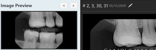
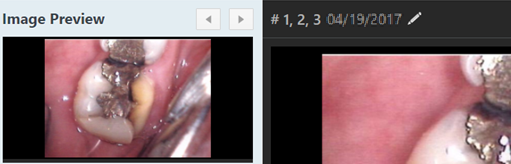
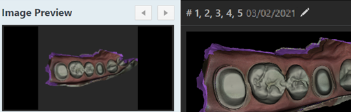
For an individual intraoral X-ray, individual intraoral photo, CAD/CAM scan, or 3D snapshot, under Image Preview, there is only one thumbnail image. A larger version of the image displays in the viewing area.
-
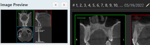
For a 3D volume, under Image Preview, there is a thumbnail image of an axial slice of the volume. The Coronal, Sagittal, and Axial views of the volume display in the viewing area.
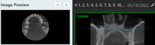
-
For a series of intraoral X-rays or a set of intraoral photos, under Image Navigator, there are multiple thumbnail images. A larger version of the selected image (as indicated by the yellow box around the thumbnail) displays in the viewing area.
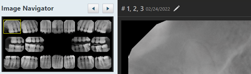
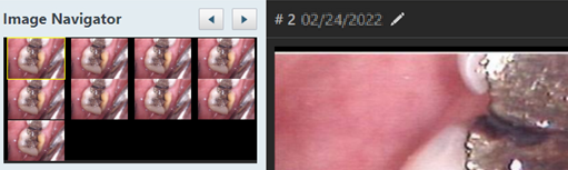
-
-
If you are viewing a series of intraoral X-rays or a set of intraoral photos, under Image Navigator, clicktap the thumbnail image that corresponds to the image for which you want to assign or change tooth numbers; otherwise, skip this step.
-
ClickTap the Edit
 icon.
icon. -
When prompted with the Assign Tooth Numbers window, clicktap the Image Details link.

-
Review the image details.

-
When ready, clicktap the Return to Chart link to return to the tooth chart view or clicktap the X to close the window.
Comments
0 comments
Please sign in to leave a comment.